
Retinal Dysplasia in Dogs: Folds, Geographic, and Detached Forms Explained
Few certification findings cause as much confusion as retinal dysplasia. The name sounds catastrophic, and in its worst form it is — yet the most common form is a minor blemish that never affects a dog's vision or its suitability for breeding. The difference between "ignore it" and "remove this dog from the program" comes down to one word on the exam report: the grade. Understanding the three forms of retinal dysplasia is the difference between a sound breeding decision and an avoidable mistake in either direction.
Retinal dysplasia is an abnormal development of the retina, the light-sensitive layer at the back of the eye. Instead of forming a smooth, even sheet, the retinal tissue folds, clumps, or in severe cases fails to attach properly. Because it is a developmental fault present from birth, an ophthalmologist can usually detect it in young puppies, which makes it one of the conditions worth screening for early rather than waiting for adult certification.
The Three Forms, From Mild to Severe
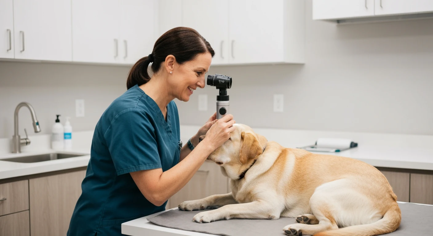
Ophthalmologists classify retinal dysplasia into three grades that reflect both severity and prognosis. They are not interchangeable, and lumping them together under one scary label is exactly the mistake that leads breeders astray.
Focal and multifocal folds. This is the mildest and by far the most common form. Small folds or wrinkles appear in the retina, visible to the ophthalmologist as short linear or comma-shaped marks. These folds usually have no measurable effect on the dog's vision. In some cases they even appear to resolve as a puppy's eye grows. For most breeds, folds are a minor finding that does not remove a dog from a breeding program on its own, though they should still be recorded and weighed alongside the rest of the dog's health picture.
Geographic retinal dysplasia. The moderate form. Here the abnormal tissue forms larger, irregular, horseshoe- or geographic-shaped lesions rather than simple folds. Geographic lesions can cause localized blind spots and carry a more guarded prognosis, because the affected area of retina does not function normally. A geographic finding warrants real caution in breeding decisions and a frank conversation with your ophthalmologist about the specific dog and breed.
Detached retinal dysplasia. The severe form, and the one that justifies the alarming name. The retina fails to attach properly and may detach partially or completely, causing serious visual impairment or total blindness in the affected eye. This form is genetically distinct in its impact and is a clear reason to remove a dog from breeding. In certain breeds it is associated with skeletal abnormalities, discussed below.
The Oculoskeletal Connection
In a few breeds — notably the Labrador Retriever and the Samoyed — the gene associated with severe retinal dysplasia is also linked to skeletal dysplasia, a condition called oculoskeletal dysplasia or dwarfism. Dogs that inherit two copies of the relevant mutation can show both the blinding retinal detachment and shortened, abnormal limbs. Dogs with a single copy may show only mild retinal folds while appearing skeletally normal. This is why a "minor" fold in one of these breeds deserves more thought than the same finding in a breed without the skeletal link: the fold may signal carrier status for a far more serious recessive condition. Where a DNA test exists for the breed-specific mutation, it can clarify what a clinical fold alone cannot.
Which Breeds Are Predisposed
Retinal dysplasia appears across many breeds, but several are recognized as predisposed and are routinely screened. The Labrador Retriever carries the oculoskeletal form noted above. The English Springer Spaniel and the American Cocker Spaniel see folds and geographic forms with some frequency. The Cavalier King Charles Spaniel and the Sealyham Terrier are also among the breeds where the condition is well documented. Predisposition does not mean every dog is affected — it means screening matters more in these breeds, and a finding should be interpreted against what is known for that specific breed. For a broader view of which conditions cluster in which breeds, see the guide to hereditary eye diseases in common breeds.
How the Grade Drives the Breeding Decision
The practical rule that experienced breeders follow is that the grade, not the diagnosis, decides the action. A multifocal fold in a breed without the skeletal link is typically recorded as a breeder-option finding and does not, by itself, end a dog's breeding career — the decision is made by weighing the fold against the dog's overall quality and the partner it is paired with. Geographic lesions raise the bar considerably and demand caution. The detached form is a straightforward removal from breeding.
This tiered approach is exactly what the breeder-option system was designed to support. Rather than a blanket pass or fail, it gives breeders the information to make a graduated decision. The logic of working with these findings rather than panicking over them is covered in the guide on breeder-option eye certification findings, which is essential reading for anyone whose dog comes back with a non-clear result.
In Labradors and Samoyeds, even mild retinal folds can indicate carrier status for the recessive oculoskeletal mutation. Pairing two such carriers risks producing affected puppies with both retinal detachment and dwarfism. In these breeds, treat any fold as a reason to choose a clear or DNA-tested partner, not as a finding you can safely double up on.
What to Confirm on the Exam Report
When your dog's eye exam returns a retinal dysplasia notation, do not stop at the word itself. Confirm the form — folds, geographic, or detached — because that single distinction changes everything that follows. Ask whether it is unilateral or bilateral. In predisposed breeds, ask your ophthalmologist whether a DNA test is available to clarify carrier status. And record the finding accurately in your own files so it travels with the dog and informs the next generation's pairings. Eye health managed across generations, rather than one exam at a time, is what genuinely reduces these conditions in a line — the principle explored in the guide to eye certification requirements by breed.
Retinal dysplasia is a perfect example of why breeders should understand findings rather than fear them. The same two-word diagnosis can describe a harmless wrinkle or a blinding malformation. The grade tells you which one you are dealing with — and an informed breeder who reads that grade correctly makes better decisions than one who panics at the name.